How do complex neural connections give rise to cognition and behavior, and how do brain disorders alter these processes? Using the common marmoset as our primary nonhuman primate model, we integrate neuroimaging, connectomics, spatial omics, brain–computer interfaces, and computational modeling to investigate brain organization across scales, from molecules and cells to whole-brain networks. Building on this foundation, we investigate disease mechanisms, develop digital twin models that predict brain activity and responses to interventions, and compare biological and artificial neural networks to uncover fundamental principles of intelligence. Our research focuses on three interconnected directions.
1. Neuroimaging and Brain Disorders
We develop high-resolution structural MRI, awake marmoset functional MRI, multinuclear MRI, and multimodal analytical methods to improve the characterization of fine brain structures, connectivity patterns, and dynamic activity. Focusing on brain development and neurodegenerative disorders, we establish and investigate marmoset disease models, combining longitudinal imaging with behavioral monitoring to track disease onset and progression. We further integrate brain connectivity atlases, cell-type information, and spatial gene expression data to uncover the cellular and circuit mechanisms underlying imaging phenotypes. These mechanisms are tested through pharmacological interventions and targeted neuromodulation, providing a foundation for identifying imaging biomarkers and intervention targets with translational potential.
2. Brain Atlases and Digital Twins
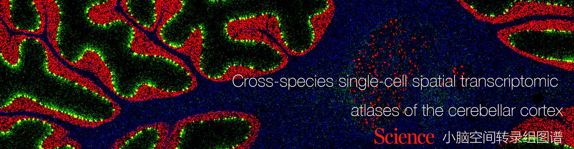
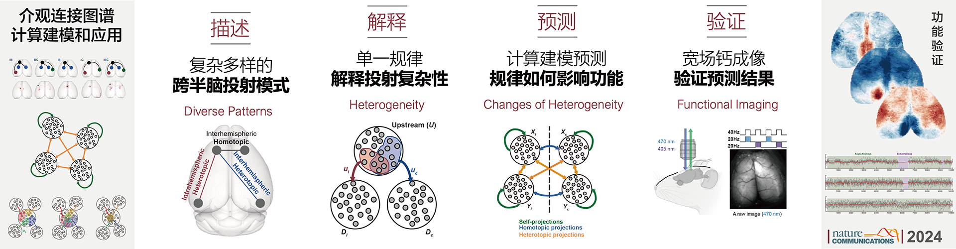
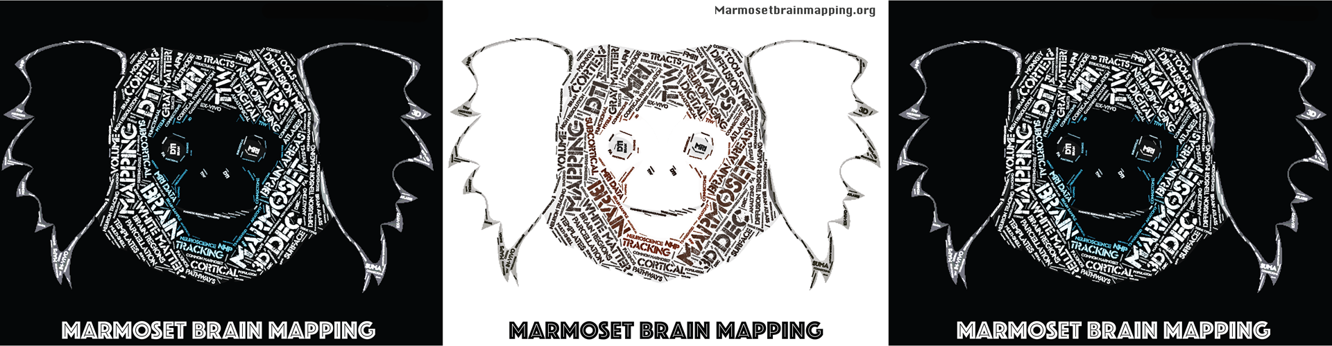
Through the Marmoset Brain Mapping Project, we have developed multimodal atlases encompassing cortical parcellations, white matter pathways, population templates, functional connectivity, and the cerebellum. By further integrating mesoscale fiber reconstruction with single-cell spatial transcriptomics, we have revealed cellular and connectivity features of the primate cerebellum, as well as opposing molecular gradients underlying cortical organization. Building on these advances, we combine functional imaging, brain–computer interfaces, and computational modeling to develop digital twins of the brain. Cross-scale atlases provide biological constraints, while whole-brain functional imaging and local neural recordings characterize dynamic activity. Controlled perturbations—including electrical stimulation, optogenetic manipulation, and pharmacological interventions—allow us to test how brain networks respond. Through iterative calibration between experiments and models, we aim to develop dynamic models that reproduce brain network activity and predict intervention responses, supporting the study of brain function, the identification of neuromodulation targets, and the optimization of intervention strategies.
3. Brain-Inspired Intelligence: Reciprocal Insights Between Biological and Artificial Neural Networks
We establish a quantitative framework for comparing biological and artificial neural networks from a connectomic perspective, examining how connectivity structure and activity correlations relate to network function. Our goal is to identify shared computational principles and organizational mechanisms unique to each system. In one direction, we apply brain network analysis to artificial intelligence, tracking how structure–function relationships evolve during training and investigating how module formation, information integration, and network architecture influence learning. These insights guide the exploration of connectivity-informed network design, training optimization, and model compression. In the other direction, we use artificial neural networks as fully observable and precisely controllable systems to test and refine connectomic methods, generating new hypotheses and principles of network learning and information processing and advancing reciprocal discovery between neuroscience and artificial intelligence.

Senior Investigator