Understanding brain function requires fathoming into the activities and self-organization of networks at different spatio-temporal scales. At the micro- and meso-scopic levels, studies use intracranial methods that can acquire and analyze signals produced by single cells or microcircuits. At the macro-scopic levels, on the other hand, studies concentrate on the activity of whole-brain networks, however based on surrogate signals, such as measurements of blood flow, volume, and oxygenation. Many of these recordings suffer by so-called ill-posed problem-nature: the measured signals can have all kind of different, and occasionally inter-contradicting natures, and surely interpretations. A dominating methodology, for example, in neuroimaging is functional MRI (fMRI), which is based on the blood-oxygen-level-dependent (BOLD) signal. While the method offers an attractive global view of brain-activation, the underpinning of such activations is always ill-defined. A highly promising method that could – together with electrophysiological measurements substantially improve imaging interpretations is so-called molecular fMRI, namely measurements of concentration changes of neurotransmitters and neuromodulators. This could be a substantial step forward in achieving precise and reliable visualization of brain activity by observing the neuronal processes on molecular and cellular level with outstanding specificity.
Molecular fMRI is a methodology that combines molecular imaging and fMRI. It allows for the direct monitoring of physiological processes within the brain with a spatiotemporal resolution that is not achievable by any other existing method. Molecular fMRI is performed with the aid of exogenous substances – MRI contrast agents, which are designed to be sensitive to various ions or molecules involved in brain physiology and produce the alteration of contrast in response to changes in their microenvironment. These functional markers are called bioresponsive or smart contrast agents (SCAs). Notably, only a handful of labs worldwide possess the appropriate expertise to design, obtain and validate the SCAs in vivo.
Bioresponsive or smart contrast agents
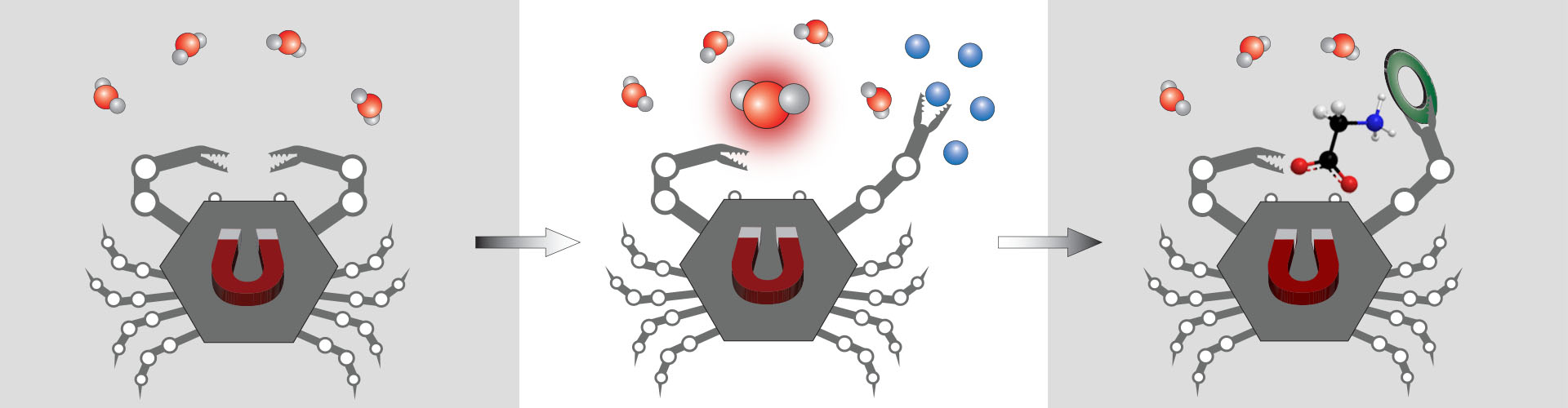
We are developing bioresponsive SCAs, the probes capable of varying the MR image contrast upon changes in the local environment. They strongly respond to different aspects of brain physiology, such as calcium ions or amino-acid neurotransmitters, thus dramatically improving the specificity of fMRI measurements. We use a multitude of strategies to prepare these SCAs and apply them by means of the cutting-edge MRI techniques that have recently been established. Besides the SCAs for conventional T1- and T2-weighted MRI, we are developing sensors suitable for fluorine-based, chemical exchange saturation transfer (CEST) or hyperpolarized MRI. To improve the biokinetic properties of these functional tracers and mitigate their use in vivo, we combine them with various nanosized and biocompatible carriers, thus obtaining multimeric, multicontrast and multimodal SCAs for molecular fMRI.
Functional MRI studies with bioresponsive contrast agents
We are also developing methods to validate this molecular fMRI approach in vivo and utilize its indefinite potential to visualize vast number of biological processes. Our goal is establishment of routine methods that assess various aspects of neural signaling with high specificity. To this end, the above described and in-house designed and prepared small- and nano-sized SCAs are used for the development of novel functional neuroimaging methods. With these, we are pursuing the following molecular fMRI studies:
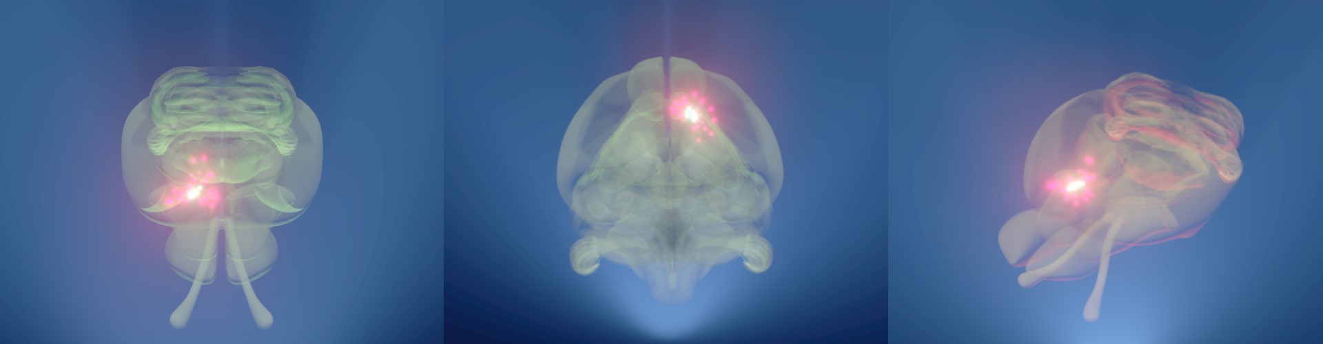
a) Dynamic fMRI recordings of extracellular calcium and amino-acid neurotransmitters. Triggering events with significant biomedical relevance are used in combination with the applied SCA to visualize various neuronal events in dynamic fashion with high spatial and temporal resolution.
b) Concentration-mapping of diverse ionic and/or molecular targets (e.g. pH or metal cations). We record multiple MRI maps in the region of SCA application by using the state-of-art MRI techniques for the quantitative assessment. This unique combination allows for the precise correlation of the recorded MRI signal with the concentration of the desired target. In turn, the quantitative molecular fMRI can substantially broaden the scope of contemporary neuroimaging techniques to study brain metabolism and function.
From the design of bioresponsive contrast agents to molecular fMRI studies
During the former period of our research at the Max Planck Institute for Biological Cybernetics, we have made a remarkable progress in the development of various types of MRI contrast agents. Altogether, we possess the largest number of the bioresponsive MRI agents that are suitable for neuroimaging studies by means of molecular fMRI.
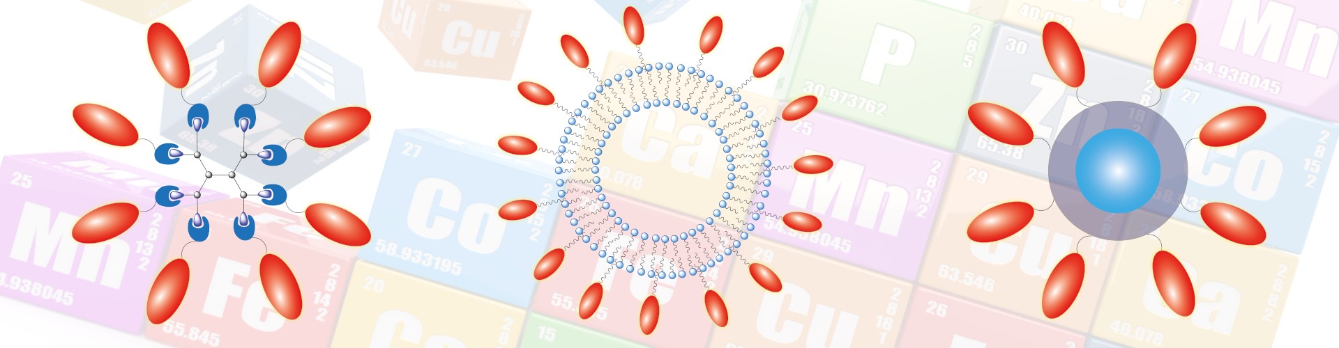
Our expertise has been diligently developed and accumulated over the years, to result in numerous examples of bioresponsive, target-specific, multicontrast and multimodal contrast agents that have been prepared, analyzed and reported. We currently have the biggest library of the Ca-sensitive MRI agents that are suitable for MRI studies at the proton and fluorine frequencies. Together with the most recently reported Zn-sensitive probe, these agents exhibit the highest MRI signal changes in vitro than any other competing probes. Additionally, we have developed a series of neurotransmitter-sensitive MRI probes that respond to a range of amino acid-based neurotransmitters, such as Gly, Asp or Glu.
In parallel, we synthetically modified our bioresponsive probes and prepared liposome-, nanoparticle- or dendrimeric-based MRI agents, which showed extremely advantageous in vitro and in vivo properties. These nanosized contrast agents display preferred biokinetic properties, i.e. have long tissue retention time, and are prone to further modifications and functional optimizations.
With these extraordinary probes in hand, we executed several studies to demonstrate their potential for functional MRI studies. Our nanoparticle-based Ca-sensitive agent was the first probe reported in the literature to show the MRI signal changes in vivo upon using calcium ions as the stimulus (Moussaron et al, Small 2015). More recently, we also utilized a Ca-sensitive MRI probe in studying brain ischemia. We showed that an ischemic stroke can be early detected and monitored by means of our molecular fMRI approach (Savić et al, PNAS 2019). These findings are extremely encouraging and show great potential for the development of numerous approaches to study the brain function at molecular and cellular level. Our lab will proceed in expanding the scope of the SCA-assisted fMRI to help understanding various physiological and pathological processes in brain, and consequently deliver new and valuable insights to biomedical research.

Senior Investigator