Time:2020-06-14
Two recent research articles revealed a considerable degree of plasticity of retinal function during the early stage of development. The research was completed by Dr. ZHANG Yifeng’s lab of the Institute of Neuroscience, Center for Excellence in Brain Science and Intelligence Technology of the Chinese Academy of Sciences.
Plasticity is essential for the normal functioning of the nervous system. When an individual develops to a certain stage, the brain begins to obtain external information from the sensory organs, and makes modifications to neuronal connections based on this information, so as to more effectively respond to external stimuli. Therefore, the neurons at this stage showed much higher plasticity. If the external environmental stimuli deviates from the normal environment during this early developmental stage, it will lead to changes in the function of the nervous system. Taking mouse vision as an example, if the eyelids are sutured before eye opening to block the input of visual stimuli, it will cause problems in the basic features of the primary visual cortex, such as the ocular dominance columns, direction selectivity and orientation selectivity; while if a rich environment is provided during the critical period of mouse visual cortex development, the establishment of functions such as the ocular dominance column can be accelerated, this may even make up for the functional defects caused by genetics, such as the topological projection defects caused by gene knockout. Previously, a fair amount of research on the development of the visual system has been carried out with changed visual environment, but the main subject of those studies has almost always been the visual cortex. Few people think that the visual sensor, the retina, will also show plasticity, i.e. changed by external stimuli. So, can retinal neurons be changed by external visual stimuli?
In this study, mice were given visual stimulation with different directions of motion from postnatal day 10 to 35. Multi-channel electrode arrays, single-channel and dual-channel whole-cell patch clamps were used to record and analyze the light response of ON-OFF direction-selective ganglion cells (ON-OFF DSGCs) in the retina, and it was found that these cells have significant changes in at least two aspects. One is that they have significantly improved ability of motion direction encoding. As long as the visual experience training contains sufficient motion stimulation, ON-OFF DSGCs show significantly improved coding ability for all motion directions. Further research found that the excitatory inputs received by ON-OFF DSGCs became more stable after training, which led to more reliable responses in these cells, thereby improving the accuracy of their coding of motion directions. Secondly, there are gap junction connections among the upward-preferring ON-OFF DSGCs, and the strength of the connections can be enhanced by the upward visual motion experience training, and weakened by the downward training. Gap junction are closely linked tosynchronized firings among connected cells. The study shows that the stronger the synchronized firing during visual motion experience training, the more obvious the improvement of the gap junction connections afterwards, this is the typical characteristics of Hebbian plasticity. Both effects can last for a long time and cannot be affected by the change of visual experience later.
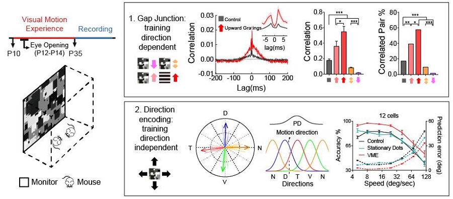
Legend: Left, Diagram of the visual motion experience (VME) training. Right (top), The percentage and strength of gap junction connections among upward preferring ON-OFF DSGCs are highly correlated with the directions of VME training. Right (bottom), improvement of motion direction encoding of ON-OFF DSGCs is independent of VME training directions. (Image by CEBSIT)
These two studies demonstrated for the first time that the response properties of retinal ganglion cells can be changed by a visual environment enriched in motion right after eye-opening, revealing a considerable degree of plasticity of retinal function during the early stage of development. The studies enriched the understanding of experience dependent plasticity, and suggested that plasticity is likely to be ubiquitous in the nervous system, including sensory organs. The research results provide a basis for exploring how environmental stimuli change the retinal circuits, and help to explore the use of early sensory experience training for adjuvant treatment of some sensory system diseases.
These two research articles entitled “Early visual motion experience shapes the gap junction connections among direction selective ganglion cells” and “Early visual motion experience improves retinal encoding of motion directions” were published on PLOS Biology and the Journal of Neuroscience on March 25, 2020 and June 12, 2020. The research was completed by Dr. ZHANG Li under the supervision of Dr. ZHANG Yifeng, and WU Qiwen also made important contributions. The research was funded by Shanghai Municipal Government.
AUTHOR CONTACT: ZHANG Yifeng
Institute of Neuroscience, Center for Excellence in Brain Science and Intelligence Technology of Chinese Academy of Sciences
320 Yue-Yang Road, Shanghai 200031, China
Email: zhangyifeng@ion.ac.cn
 附件下载:
附件下载: 
