Time:2020-11-09
A recent study published in Movement Disorders demonstrated that hyper-activation and weakened functional connectivity of the subthalamic nucleus (STN) are neural markers of sequential working memory deficits in de novo Parkinson’s disease (PD). This work was performed by researchers in Dr. YE Zheng’s Lab at the Institute of Neuroscience, Key Laboratory of Primate Neurobiology, Center for Excellence in Brain Sciences and Intelligence Technology of the Chinese Academy of Sciences, Prof. ZHANG Yingshuang’s team at the Peking University Third Hospital, and Prof. ZHOU Xiaolin’s Lab at the School of Psychological and Cognitive Sciences of the Peking University.
Idiopathic PD is the second most common age-related neurodegenerative disease. In addition to motor symptoms such as tremor, stiffness, and slowness of movement, patients with PD develop non-motor symptoms such as cognitive decline, sleep disorders, and mood disorders, significantly impacting their quality of life and functional independence. There is no cure for PD. Medications and surgeries are used to treat motor symptoms. However, current therapeutic approaches have a limited effect on improving cognitive functions in PD. Studies on the neural mechanisms of cognitive impairment in PD may lead to potential therapies for enhancing cognition, which will eventually benefit many patients with PD.
When we schedule our day, we may arrange tasks in the order they come or prioritize a task that is due first. Flexible planning depends on the ability to maintain and manipulate sequential information online. In healthy brains, a neural system for sequential working memory comprises the lateral prefrontal cortex, posterior parietal cortex, STN, globus pallidus, and thalamus. In aged brains, the prefrontal and posterior parietal cortices are over-activated, psychophysiological interactions between cortices are weakened, but functional connectivity between cortices and the striatum is strengthened. Neural compensation within the network may help keep sequential working memory strong in older adults. However, sequential working memory is significantly impaired even in patients with mild PD, which coincides with but is independent of motor symptoms.
To investigate the neural markers of sequential working memory deficits at the early stages of PD, researchers recruited 50 patients with do novo PD and 50 healthy participants. All participants completed a digit ordering task in a magnetic resonance imaging (MRI) scanner when their brain activity was recorded. In this task (Fig.A), participants were asked to remember a sequence of four digits and recall the digits in ascending order. Two memory processes were measured in the task: participants only needed to remember the original sequence if the digits appeared in ascending order (maintenance of sequences); they had to reorder the digits (manipulation of sequences) and recall the new sequence if the digits were fully randomized. Participants’ blood oxygen level-dependent (BOLD) functional MRI signals were recorded when they performed the task. The increase/decrease of the BOLD signals reflects the increase/decrease of regional brain activity.
Researchers analyzed regional brain activations and inter-regional functional connectivity in the neural network for sequential working memory. They found that patients’ poor performance correlated with hyper-activation of the STN (Fig.B) and weakened functional connectivity between the STN and striatum (Fig.C). Healthy participants who showed lower STN activation and more robust STN-striatum connectivity performed more accurately in maintaining sequences. The STN was more activated, and the task accuracy was decreased when participants had to reorder the digits. Healthy participants who showed greater ordering-related STN activation change exhibited smaller accuracy cost in manipulating sequences. That means the STN may play a modulatory role in maintaining and manipulating sequential information. The STN’s modulatory role was remarkably weakened in patients with de novo PD. The modulation was partially realized by the STN-striatum functional connectivity. The contribution of the STN activation or activation change was absent.
This work demonstrated STN’s modulatory role in sequential working memory and linked sequential working memory deficits in de novo PD with STN’s altered functions. It implies that down-regulation of the STN activation and up-regulation of the STN functional connectivity may be a potential strategy for enhancing PD’s sequential working memory.
This work entitled “The role of the subthalamic nucleus in sequential working memory in de novo Parkinson’s disease” was published online in Movement Disorders on October 24, 2020. Dr. YE Zheng at the Institute of Neuroscience, Center for Excellence in Brain Sciences and Intelligence Technology of the Chinese Academy of Sciences, is the first author and co-corresponding author. Prof. ZHANG Yingshuang at the Peking University Third Hospital and Prof. Thomas Münte at the University of Lübeck are co-corresponding authors. ZHANG Guanyu at the Chinese Academy of Sciences Insitute of Psychology and LI Shuaiqi at the School of Psychological and Cognitive Sciences of the Peking University contributed to this work. This work was supported by the National Natural Science Foundation of China, the German Research Foundation, and the Alexander von Humboldt Foundation.
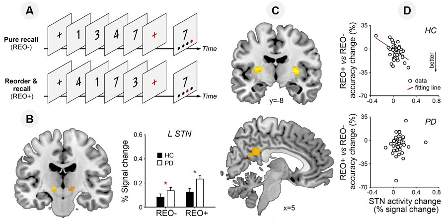
Figure legend: (A) Digit ordering task. (B) Hyper-activation of the subthalamic nucleus (STN) in de novo PD. (C) The STN functional connectivity with the putamen and other brain regions are weakened in de novo PD. (D) The modulatory role of the STN is weakened in de novo PD. (Image by CEBSIT)
AUTHOR CONTACT:
YE Zheng
Institute of Neuroscience, Center for Excellence in Brain Sciences and Intelligence Technology, Chinese Academy of Sciences, Shanghai, China.
E-mail: yez@ion.ac.cn 附件下载:
附件下载: 
