Time:2025-04-30
Over the past decade, intensive studies have shown that the brain meningeal lymphatic system acts as the brain’s “waste disposal network”, maintaining homeostasis by clearing metabolic waste and transporting immune cells.
However, the mechanism underlying its development regulation remains unknown—how does this intricate system form, and which cells or signals govern its specific spatial arrangement in the meninges?
This work, led by Dr. Jiulin Du’s lab at the Institute of Neuroscience, Center for Excellence in Brain Science and Intelligence Technology, Chinese Academy of Sciences, uncovers the core regulatory mechanism of brain meningeal lymphatic system development.
Taking advantage of in vivo long-term imaging in zebrafish, in combination with genetic manipulations and neural activity studies, the research team found that increasing neural activity (such as visual stimulation) significantly enhances the development of muLECs in the leptomeninges, while inhibiting neural activity (such as visual deprivation) leads to a significant reduction in the muLEC number. Focusing on Vegfc, a key factor necessary for lymphatic system development, the team identified a specific glial subpopulation—slc6a11b+ RAs—that expresses Vegfc (Figure I). These cells extend fibers to the brain surface, making them the main source of Vegfc in the brain. Deletion of slc6a11b+ RAs impairs muLEC development, while upregulating these cells’ Vegfc signaling enhances muLEC development (Figure II). Furthermore, slc6a11b+ RAs’ activity and their Vegfc expression levels are positively regulated by neural activity (Figure III). The team further discovered that the precursor Vegfc (pro-Vegfc) secreted by slc6a11b+ RAs requires the cooperation of ccbe1+ fibroblasts to be converted into its mature form (mVegfc). This cross-tissue collaboration precisely restricts the distribution of mVegfc to the brain-meningeal interface, ensuring that lymphatic endothelial cells are confined to the brain surface, preventing their invasion into the brain parenchyma, which could cause immune disruptions (Figure IV).
The study reveals that the brain not only serves as the center for neural information processing but also acts as a “coordinator” of its microenvironment. Neural activity dynamically regulates the development of brain lymphatic endothelial cells through specific glial subpopulations, revealing a novel "neural-glia-fibroblast-lymphatic" regulatory axis. This provides a new framework for understanding how the brain adapts its lymphatic network based on functional needs, and it also explains why the brain meningeal lymphatic system remains confined to the meninges, avoiding invasion into the brain parenchyma. In the future, intervening in this regulatory network may offer new perspectives for understanding the role of the meningeal lymphatic system in neurological diseases such as neurodegenerative disorders and potential strategies for developing treatments.
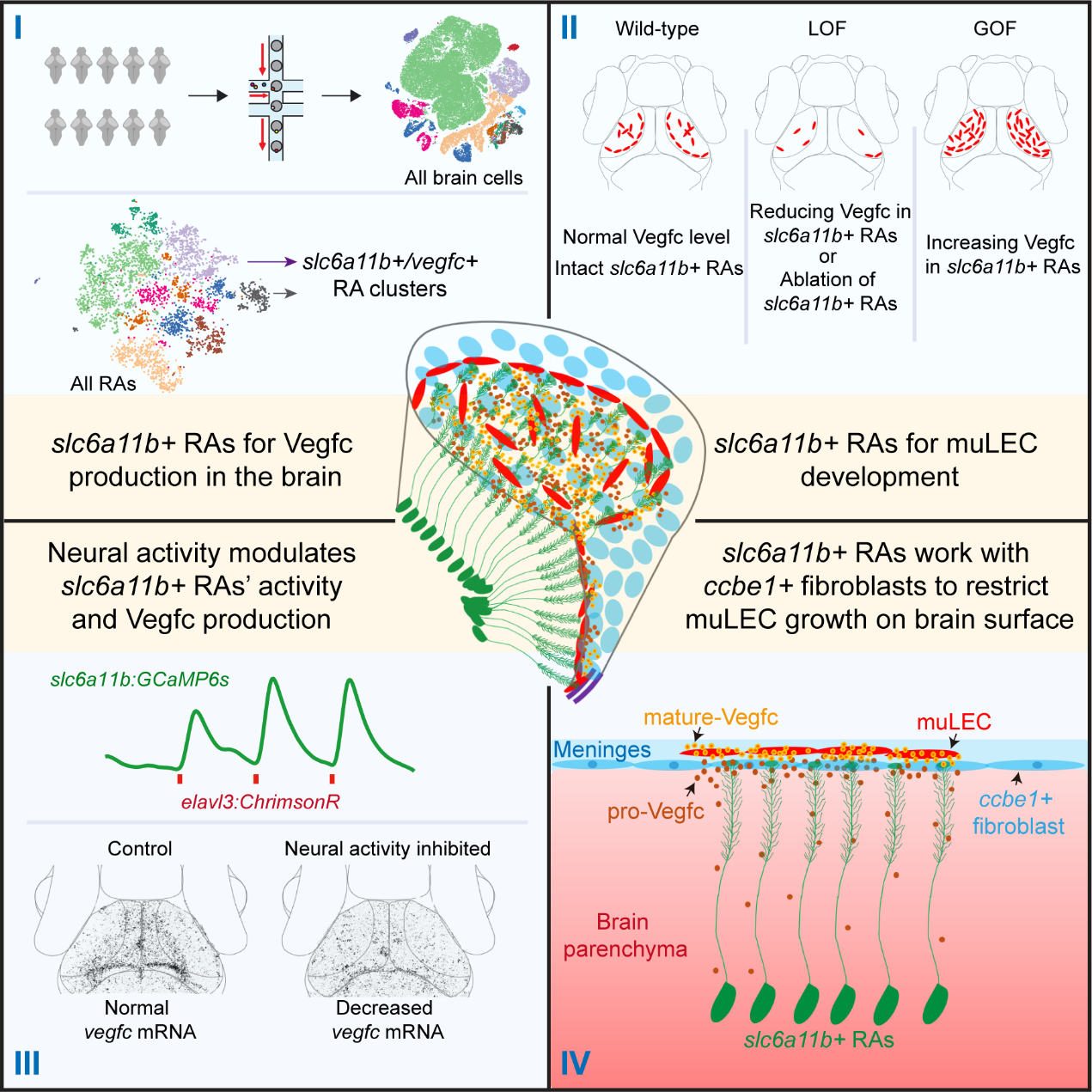
Figure I- Figure IV
Figure I. slc6a11b+ RAs are the primary source of Vegfc in the brain
Figure II. Vegfc produced by slc6a11b+ RAs controls muLEC development
Figure III. Neural activity modulates slc6a11b+ RAs’ activity and their Vegfc production
Figure IV. slc6a11b+ RAs work with ccbe1+ fibroblasts to restrict muLECs growth on brain surface
Contact:
DU Jiulin
Center for Excellence in Brain Science and Intelligence Technology, Chinese Academy of Sciences, Shanghai, China.
forestdu@ion.ac.cn
 附件下载:
附件下载: 
