Time:2021-04-10
In a study published in eLife by Dr. GU Yong’s lab (the lab of Spatial Perception) at the Institute of Neuroscience, Center for Excellence in Brain Science and Intelligence Technology of the Chinese Academy of Sciences, they used a virtual reality system in conjunction with extracellular electrophysiological recording techniques in awake macaques to investigate the encoding of self-motion signals in posterior cingulate region neurons in the macaque brain, and found that posterior cingulate cortex (PCC) carries robust vestibular signals with complex temporal-spatial tuning properties.
In cognitive studies, landmark-based and vector-based navigation are two commonly used strategies for spatial tasks. In the absence of effective external landmarks such as in open fields of deserts and grasslands, organisms mainly rely on vector-based navigation (also known as path integration) to explore the world, in which self-motion cues from multiple sensory channels such as optic flow and vestibular ones for updating and accumulating one’s heading direction and location are used. As the well-known navigation system in the animal brain, the hippocampus system plays a key role in path integration. However, it is not yet understood how the hippocampus uses external self-motion information to update one’s heading direction and location in path integration. In previous studies, encoding of vestibular and optical flow visual signals has been identified in several sensory brain areas in the macaque brain (mainly located in the temporal and parietal lobes), however, these regions have no direct anatomical connection to the hippocampal system.
Previous studies have shown that the posterior cingulate region, located above the corpus callosum, is structurally connected with both the self-motion-related sensory cortex and the hippocampal system. Therefore, to find the brain regions that play a bridging role in the transmission of self-motion information to the navigation system, researchers in GU Yong 's group used a sophisticated virtual reality system with a six-degree-of-freedom motor platform to investigate the representation of self-motion information in the anatomical hub in this pathway-the posterior cingulate region. In the experiments, 26 different directions of motion stimuli almost uniformly distributed in three-dimensional space were given by the platform, and visual stimuli were simulated with optic flow.
The peripheral vestibular system of an organism consists of two main parts: the otolith which encodes linear acceleration (translational signals) and the semicircular canal, which encodes rotation. Thus, motion stimuli were given in both translational and rotational conditions. By extracellular recordings in the two different subregions of three macaques-posterior cingulate cortex (PCC) and retrosplenial cortex (RSC), the researchers found that the posterior cingulate cortex contains robust vestibular signals, which have complex temporal and spatial tuning properties. In contrast to PCC, RSC, which is deeper in the brain and also encodes vestibular signals, do not have a clear temporal and spatial tuning property.
To quantify this temporal and spatial tuning property, they decomposed the temporal and spatial components of motion stimuli by a three-dimensional temporal-spatial dynamic model, and found that PCC neurons carry multiple temporal components, such as acceleration, velocity, jerk, and position, indicating that acceleration signals from peripheral vestibular organs are integrated or differentiated in different degrees during transmission to this brain region to be used for different functions. In addition, neurons here carry more acceleration vestibular signals in translational movements, while the encoding in rotational condition is more velocity based. Considering the distribution of preferred directions of neuron population in rotation, the vestibular signals here are likely to involved in head direction cells in the hippocampal system. In contrast to the strong vestibular signals here, the visual information in both regions is fairly weak, implying that this region may not integrate both types of self-motion information for further functions.
In conclusion, this work fills the gap in the encoding of vestibular information in the whole brain, for the discovery of another brain area in the big network . On the other hand, it provides the foundation of further exploration in the transmission of vestibular information from the sensory cortex to the hippocampal navigation system for other researchers.
This work entitled “Robust vestibular self-motion signals in macaque posterior cingulate region” was published online in eLife on April 8th, 2021. LIU Bingyu is the first author, Dr. GU Yong is the corresponding author and TIAN Qingyang also contribute to data collection and analysis. This study was supported by grants from National Nature Science Foundation of China, CAS and Shanghai Municipal Science and Technology Commission.
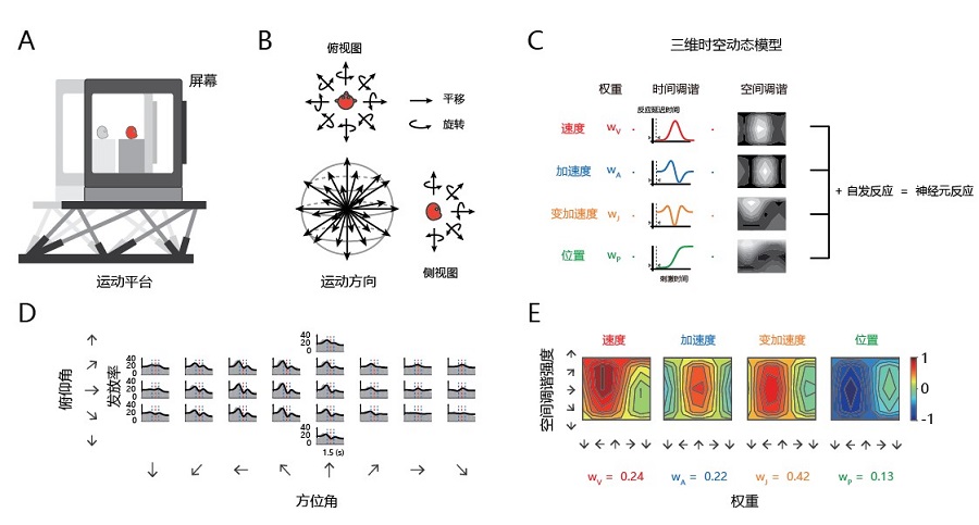
Figure: (A) Experimental setup: the virtual-reality system and the 6-degree-of-freedom motion platform. (B) Illustration of 26 translational motion directions which are equally distributed in the three-dimensional space and the corresponding 26 rotational stimuli. (C) Illustration of the 3D temporal-spatial dynamic model. (D) An example neuron which responded to vestibular stimuli in translational condition. The gray areas show neural responses, and the black lines indicate model fitting results. (E) The spatial kernel and the weight of each temporal component after model fitting. (Image by CEBSIT)
AUTHOR CONTACT:
GU Yong
Center for Excellence in Brain Science and Intelligence Technology , Chinese Academy of Sciences, Shanghai, China.
E-mail: guyong@ion.ac.cn
 附件下载:
附件下载: 
