Time:2017-12-20
A recent study published in PNAS provides a precise 3-D whole-brain atlas for the cholinergic system and the complete morphology of individual cholinergic neuron in the basal forebrain. The research was performed by researchers in Dr. LUO Qingming and Dr. GONG Hui’s team at Huazhong University of Science and Technology and Dr. QIU Zilong’s Lab at Institute of Neuroscience, Chinese Academy of Sciences. This work provides a valuable approach for classifying neuronal cell types based on their genetic labeling, connectivity, and morphology.
The cholinergic system in the brain plays crucial roles in regulating sensory and motor functions as well as cognitive behaviors by modulating neuronal activity throughout the cortex and subcortical nuclei. Dysfunctions of cholinergic neurons can lead to neural disease such as Alzheimer disease and sleeping disorders. Generation of a whole-brain atlas for the cholinergic system will be essential for understanding the functions of cholinergic neurons. However, previous studies based on 2-D sectioning and immunostaining can only provide the rough distributions and estimated number of cholinergic neurons. The precise 3-D whole brain atlas of cholinergic neurons is still lacking. Moreover, since the lack of precise annotation and accurate identification of cholinergic neurons with their terminal arborizations, how cholinergic neurons innervated different brain regions is still in the mist.
In order to generate the precise anatomical information of the individual neuron in the whole brain, the researchers took advantage of the newly developed transgenic mice (crossing Chat-ires-Cre mice with Ai47 reporter mice) and virus labelling technique to specifically label the cholinergic neurons in mouse brain. Using a recently developed whole brain imaging system called brain-wide positioning system (BPS) of the VBN team led by Dr. LUO Qingming and Dr. GONG Hui at Huazhong University of Science, the researchers revealed stunning neuronal distribution and morphologies of several representative populations of cholinergic neurons. A comprehensive whole-brain 3-D atlas of cholinergic neurons that contains the information of the precise location, numbers, density and soma volumes of cholinergic neurons in specific brain areas were acquired. The morphology of 50 cholinergic neurons in the basal forebrain, reconstructed with the high-resolution imaging of dendritic and axonal processes, showed that adjacent cholinergic neurons can project to multiple distinct nuclei, which is in contrast to previous hypothesis. The accurate identification and anatomical location of the terminals and the projected areas indicated a new organized model of cholinergic projections, in which individual cholinergic neuron tends to project to interconnected brain areas. This 3D atlas may shed light on brain disorders associated with cholinergic system in the brain and spark inspiration for cholinergic neuron classifications.
This work entitled “Generation of a whole-brain atlas for the cholinergic system and mesoscopic projectome analysis of basal forebrain cholinergic neurons” was published online in PNAS on December 19, 2017. Dr. LI Xiangning from VBN team and graduate student YU Bin from Institute of Neuroscience are the first authors with equal contribution. Dr. Linda Madisen and Dr. ZENG Hongkui at Allen brain institute also contributed to this work. This work was supported by the National Key Scientific Instrument & Equipment Development Program of China (2012YQ030260); the Science Fund for Creative Research Group of China (61421064); National Natural Science Foundation of China Grants 91432105, 91432111, 81527901, and 31625013; and the Director Fund of Wuhan National Laboratory for Optoelectronics.
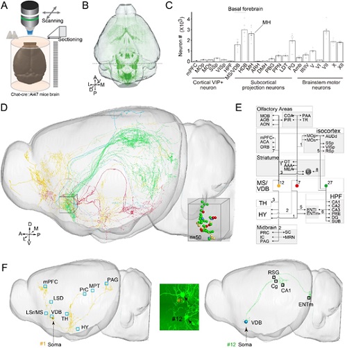
Figure legend: (A) Brain-wide positioning system (BPS). (B) Horizontal view of genetically labeled cholinergic neurons in the whole brain. (C) Numbers of cholinergic neurons in brain regions of a single hemisphere. (D) A total of 50 cholinergic neurons in basal forebrain were reconstructed from the whole-brain database. (E) Schematic diagram illustrating the major projection patterns of the cholinergic neurons in VDB. (F) 3D view of the different projection pattern of adjacent neurons (#1 and #12).
 附件下载:
附件下载: 
