Time:2017-02-08
A recent study published in Molecular Psychiatry demonstrated that autism-associated Dyrk1a truncations inhibit neural dendritic and spine growth and interfere with cortical migration during development. This work was performed by researchers in Dr. WU Bailin’s team in Children’s Hospital of Fudan University and Dr. QIU Zilong’s Lab at the Institute of Neuroscience, Chinese Academy of Sciences. This work detected 9 missense variants on DYRK1A in autism patients, and then unraveled the role of DYRK1A and the variants in the process of neuronal development. The results showed that Dyrk1a played a critical role in neural development, furthermore, two truncated mutations (R205X and E239X) led to loss-of-function of DYRK1A protein. This work reveal the neuronal functional role of autism-related DYRK1A variants for the first time, and provided a new path to learn DYRK1A protein and pathogenesis of autism better.
Autism is a complex heterogeneous disorder and belongs to the neurodevelopmental disorders. It is defined by clinical assessment and onset of three core disturbances before three-year old: impaired social interaction, repetitive-restrictive behaviors, and language abnormalities. The treatment of autism and the establishment of autism animal model are critical goals in both medicine and neuroscience.
Previous studies have suggested that the factors leading to the occurrence of ASD were very complex, including the copy number variants (CNV) in human genome or the common mutation and rare mutation on the single genes. About hundreds of genes were reported to be associated with ASD, but only dozens of them were confirmed.DYRK1A gene on Chromosome 21 that was well known for its contribution to Down’s syndrome was also one of the candidate genes of autism. Recently, variants on DYRK1A began to be detected in autism patients through exome sequencing and the association between DYRK1A gene and autism need to be examined immediately.
In order to investigate the role of DYRK1A protein as well as the variants related to autism(Figure a),the researchers expressed the wide type(WT)、Dyrk1a_shRNA and mutants of Dyrk1a in cultured cortical neuron (Figure b, c), rat hippocampus neuron (Figure d) and cortex of embryonic mice (Figure e, f) respectively, and then analyzed the development of neurons.
This study revealed that Dyrk1a played an important role in neural development. The imbalance of the Dyrk1a dosage affected dendrite development and axon length(Figure b, c). Consistent results were found in primary cultured mouse cortical neuron and rat hippocampal slice cultures. The three-missense mutants of Dyrk1a exhibited the same phenotype as Dyrk1a WT form. However, the two DYRK1a nonsense truncations (R205X and E239X) did not affect neurite out growth as the WT form of DYRK1a did (Figure b, c). In addition, both up- and down-regulation of Dyrk1a reduced the dendritic spine density in rat hippocampus neurons, indicating that the exact expression of Dyrk1a was necessary for neural development. The truncation mutant R205X showed the same level of spine development as the control group (Figure d), which led to the conclusion that the nonsense mutations caused a loss-of-function of the DYRK1A protein, which had effects on dendritic outgrowth in neurons, dendritic spine development and embryonic cortical migration. More importantly, cortical migration during the mouse embryonic stage was also altered by the overexpression of WT Dyrk1a from E14.5 to P0. These results demonstrated that Dyrk1a played a key role in embryonic development(Figure e).
This work also showed that Dyrk1a strongly affected various aspects of neuronal development, such as dendritic outgrowth, dendritic spine density, and cortical migration, during early postnatal development. Importantly, R205X and E239X, two DYRK1A truncations that were identified in ASD/ID/DD patients, functioned as loss-of-function mutants. These findings could provide a new path to learn both DYRK1A protein function and its specific role in the pathogenesis of autism better.
This work entitled “Autism-associated Dyrk1a truncation mutants impair neuronal dendritic and spine growth and interfere with postnatal cortical development” was published online in Molecular Psychiatry on Feb 2017. This work was supported by the MoST 973 Programs (2013CB945404, 2010CB529601 and 2011CBA00400) and CAS Strategic Priority Research Program (XDB02050400).
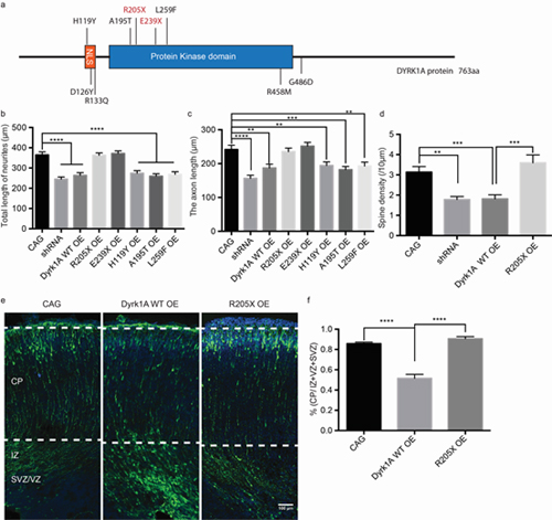
Figure legend. (a) Representation of the specific locations of missense variants related to autism in the DYRK1A protein structure, with the two truncations highlighted in red. (b-c) The statistical results for the total neurites (b) and axon length (c) between specific cell types. The bar graph shows the mean value ± SEM; the unpaired t test statistical method was used. ** p<0.01, *** p<0.001,**** p<0.0001. (d) Statistical analysis of the spine density (/10 μm) in specific types of neurons. (e-f) Dyrk1a WT over expression delayed neuronal migration in mouse embryonic cortical development. (e) Cortical migration in each condition, including CAG (left), Dyrk1a WT overexpression (middle) and R205X (right). (f) Quantification of (e).
 附件下载:
附件下载: 
