Time:2016-09-30
Researchers from the Laboratory of Brain Imaging, at the Institute of Neuroscience, CAS,revealed dynamic changesof brain connectomics for inherent functional flexibility, in which dissociablechanges of frontal and parietal cortices occurred across the human life span, using a Shannon entropy-based method.
Humans are unrivaled in their capacity to adaptively implementa wide variety of goal-directed tasks. Recent research has demonstrated that the human capability foradaptive task control is intimately related to the flexible operation of frontoparietal network. However, it remains unclear whether this flexibly functional reconfiguration is intrinsic and occurs in the absence of an overt task. To this aim, researchers proposed a probabilistic framework in which dynamic reconfiguration of intrinsic functional connectivity between brain regions can be represented as a probability distribution (Panel A). A complexity measurement (i.e., entropy) was then applied to quantify functional flexibility, the heterogeneity of dynamic connectivity between a particular region and others over time. They identified regions showing high flexibility mainly in the higher-order association cortices (e.g., LPFC, lateral parietal cortex, and lateral temporal lobes). In contrast, primary sensory (e.g., visual and auditory) areas exhibited low flexibility(Panel B). Using multiple regression analysis, they further found that flexibility of the right LPFC improved during maturation and reduced due to normal aging, with the opposite occurring for the left lateral parietal cortex(Panel C).This study not only provides a new framework toquantify the spatiotemporal characteristics of functional brain connectomics, but also sheds light on the organizational principle behind changesin brain function across the human life span.
This work entitled"Dissociable Changes of Frontal and Parietal Cortices inInherent Functional Flexibility across the Human Life Span”was published online inthe Journal of Neuroscience on Sept. 28. This work was completed by Dr. YIN Dazhi, under the supervision of Profs. WANG Zheng and CHENG Wenhong, and funded by StrategicPriority Research Program (B) of the CAS(XDB02050006), Shanghai Education Committee(HJTY2012-A06),NSFC(81471651,81571300), and Shanghai Institute for Biological Sciences, CAS(2014KIP206).
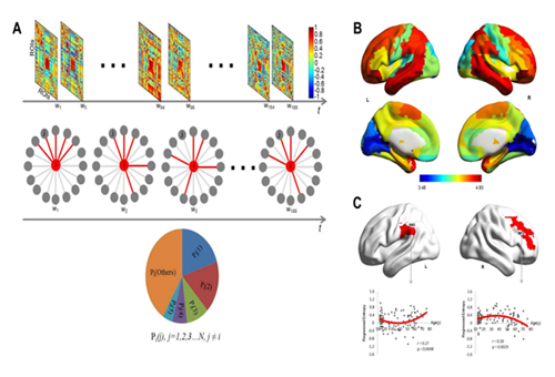
Figure legend:(A) Illustration of the probabilistic model.Dynamic functional connectivity matrices were firstly obtained for each participant using a sliding window approach.Subsequently, a local thresholdingmethod was used to reserve its k(e.g., k =5 shown here) strongest functional connections (red lines) for a given region at each time window. Then, a probability distribution Pi( j…n)was obtained, reflecting the frequency of each connectionwith i emerged across the temporal windows. A complexity measure (i.e., entropy Hi) was finally applied to this probability distribution to quantify functional flexibility of region i. (B) Brain map for inherent functional flexibility. The color bar indicates the degree of flexibility.Red colors denote high flexibility and blue colors denote low flexibility. (C) Age-related changes of functional flexibility in the left supramarginal gyrus (SMG) and right middle frontal gyrus (MFG).
 附件下载:
附件下载: 
