Time:2013-12-23
Fast reaction is essential for the survival of animals, such as when detecting and fleeing a predator. Humans or animals react rapidly in a vigilant state. The vigilance level of animals varies with brain state, which can be broadly classified into a desynchronized state dominated by small-amplitude, high-frequency activities, and a synchronized state dominated by large-amplitude, low-frequency fluctuations. To understand the neural substrate for fast reaction in the vigilant state, it is important to study brain state-dependent temporal processing for sensory cortical neurons, which are critical and early components of the sensorimotor pathway.
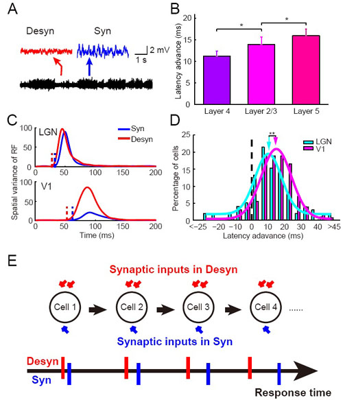
Recently, a research team (WANG Xudong, CHEN Cheng, and ZHANG Dinghong) from the laboratory of Dr. YAO Haishan at the Institute of Neuroscience, Shanghai Institutes for Biological Sciences showed that V1 neurons exhibit shorter response latency in the desynchronized brain state than in the synchronized state.
To investigate the underlying mechanism, they have measured both the resting and the visually evoked synaptic conductances of the same V1 neuron undergoing the two brain states and revealed an increase of conductance in the desynchronized state. Using a single neuron model, they showed that the conductance increase of a single V1 neuron alone is not sufficient to account for the latency advance measured experimentally. By simultaneous recording in LGN and V1, they further revealed that LGN neurons also exhibited latency advance, but with a degree smaller than that of V1 neurons. Furthermore, latency advance in V1 increased across successive cortical layers. Thus, latency advance accumulates along various stages of the visual pathway, likely due to a global increase of membrane conductance in the desynchronized state.
This research provided the first mechanistic explanation for state-dependent fast neural processing. The paper entitled “Cumulative latency advance underlies fast visual processing in desynchronized state” was published online in PNAS on December 17, 2013. The work was supported by a grant from 973 program.
Online download link: http://www.pnas.org/content/early/2013/12/17/1316166111

A. Switch of brain states indicated by changes in LFP trace. Insets above, magnified LFP traces of 4-s recording from a period in the desynchronized (marked by the red arrow) and the synchronized (marked by the blue arrow) state, respectively.
B. Latency advance increased across V1 layers. *P < 0.05, Wilcoxon rank sum test.
C. Time courses of spatial variance of RF for an example pair of simultaneously recorded LGN and V1 neurons in synchronized (blue) and desynchronized (red) states. Vertical dashed lines denote latencies of the RFs.
D. Distribution of latency advance for LGN (cyan) and V1 (magenta) neurons. Arrows point to the mean latency advance (10.4 ms for LGN; 14.4 ms for V1). **P < 0.01, Wilcoxon rank sum test.
E. A schematic model in which brain state-dependent increase in membrane conductance for cells along the visual pathway can cause cumulative latency advance.
 附件下载:
附件下载: