Time:2013-07-03
On June 28, 2013, a research article entitled "Axon position within the corpus callosum determines contralateral cortical projection" was published online in PNAS. This work was mainly carried out by graduate student Jing Zhou under the supervision of Dr. Mu-ming Poo, in collaboration with Dr. Linda J. Richards from the Queensland Brain Institute in Australia.
Corpus callosum (CC) is a large commissural tract that connects two cerebral hemispheres of the mammalian brain. Callosal axons originating from neurons of layer II/III and V of the neocortex project orderly to homotopic contralateral cortices, a process essential for bilateral coordination motor and somatosensory functions. How developing CC axons achieve this orderly projection remains unclear. Zhou et al. found that axon position within the CC plays a dominant role in determining the order of early contralateral projection.
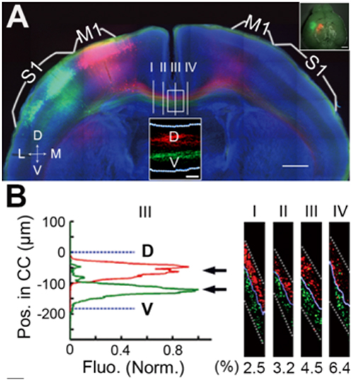
By labeling callosal axons from two nearby regions of mouse primary motor cortex (M1) and somatosensory cortex (S1), they found that callosal axons from adjacent cortical regions were orderly segregated within the CC, with those from more medial cortices locating more dorsally in the CC and projecting to more medial regions of contralateral cortices after crossing the midline.
The normal axonal order within the CC was grossly disturbed when semaphorin3A (Sema3A) / neuropilin-1(Nrp1) signaling was disrupted. However, the order in which axons were positioned within the corpus callous still determined their contralateral projection, causing a severe disruption of the homotopic contralateral projection that persisted at P30, when the normal developmental refinement of contralateral projections is completed in wild-type mice.
Their study demonstrates that the orderly positioning of axons within the CC is a primary determinant of how homotopic interhemispheric projections form in the contralateral cortex. This work was supported by 973 Program.

A, Axons from M1 and S1 are segregated in the CC. B, Fluorescence profiles of labeled axons at section III at various positions along the D-V axis of the CC.
 附件下载:
附件下载: