Time:2016-07-26
On July 26, 2016, Prof. DU Jiulin’s research group at the Institute of Neuroscience, Chinese Academy of Sciences, published an invited commentary paper entitled “A Death Trap for Microglia” in Developmental Cell to recommend two research articles published in the same issue of Developmental Cell and the latest issue of Cell Reports, respectively, both of which investigated the mechanism underlying the microglial colonization of the brain.
Microglia are the only resident immune cells in the central nervous system (CNS). In the past decade, researchers have revealed that the “resting” (inactivated) microglia in the normal brain are not resting, but continuously survey surrounding neural parenchyma through highly dynamic extension and retraction of their processes. Accumulating evidence has revolutionized our understanding of this unique cell population: microglia act not only as immune system guardians of the CNS but also as versatile sculptors for the normal development and functions of the brain, and deficiency in microglial functions is closely linked to both the generation and development of multiple brain diseases.
Microglial cells are not born in the CNS, but derive from erythromyeloid precursors (EMPs) present in the yolk sac and immigrate into the CNS from periphery during embryogenesis. How microglia colonize the CNS had remained unresolved for a long time due to technique challenging. Take advantage of the non-invasive long-term time-lapse imaging of larval zebrafish, Prof. WEN Zilong’s lab at the Hong Kong University of Science and Technology and Prof. PERI Francesca’s group at the European Molecular Biology laboratory in Heidbelberg Germany published independently two exciting studies in the latest issue of Developmental Cell and Cell Reports, respectively, demonstrating neuronal programmed cell death (PCD) during early brain development induces the entry of peripheral microglial precursors into the brain. They found that up-regulating and down-regulating of the level of neuronal apoptosis in the brain could accordingly increase and decrease the number of invading microglia (Figure 1). Furthermore, the lysophosphatidylcholine (LPC), an apoptotic cell-secreted phospholipid which was most likely released by apoptotic neurons, was shown to act as a chemoattractant to attract microglial precursor to immigrate into the brain. The two studies not only elucidate the mechanism underlying microglial colonization of the brain, promoting the understanding of early establishment of neuroimmunity, but also reveal the payoff of neuronal PCD, usually considered as an uneconomic physiological process.
Using zebrafish as animal model, Prof. DU Jiulin’s laboratory has long been engaging in investigating the neural basis for animal behavior, and has also been exploring interdiscipline research about neuro-vascular and neuro-immune interactions. As for neuro-immune interaction, his laboratory discovered that the “resting” microglia can dynamically regulate neuronal activity (Li et al., Developmental Cell, 2012). This study is recognized to be a groundbreaking work for understanding the physiological function of microglia and provides new perspective on neural-immune interactions. This study has been cited by almost all the related important review papers in the field, and at the end of 2013 it was selected as the most influential works (ranking the first) by Neuron on the journal 25-year ceremony.
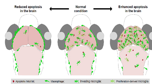
Figure1. Developmental Neuronal Apoptosis Induces Microglial Colonization of the Brain.
 附件下载:
附件下载: 
