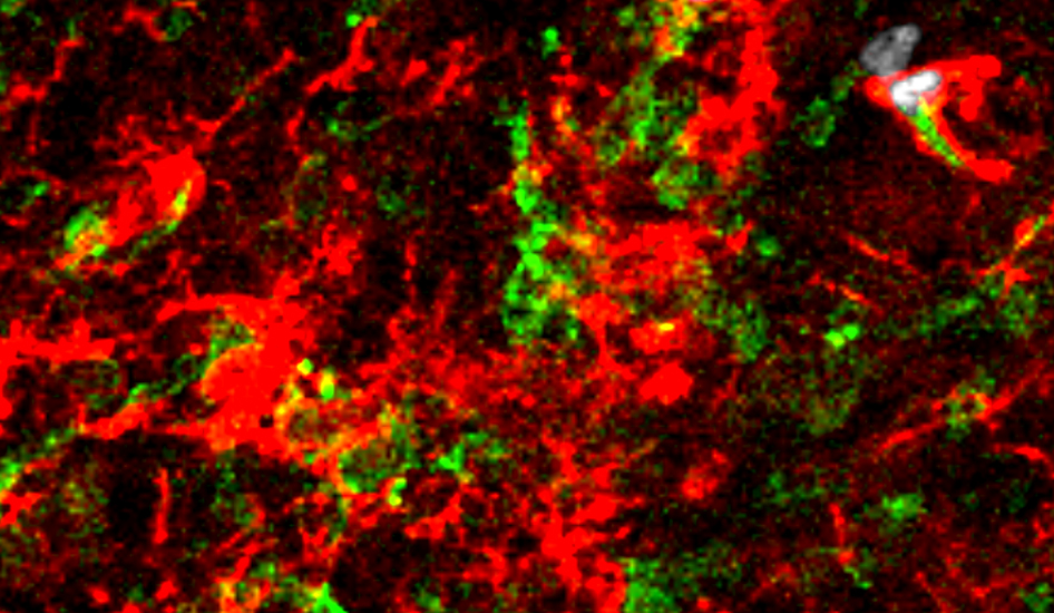
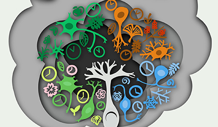
A research team led by Dr. ZHOU Jiawei found that Cspg4high microglia is a new cell source for microgliosis in neurodegeneration. They unraveled the molecular characteristics and functions of Cspg4high microglia which showed high capability of cell proliferation in neurodegenerative diseases thus providing a new insight into the pathogenesis of neurodegenerative diseases.
A recent study published in Immunity demonstrated that intestinal epithelial DRD2 specifically promotes Experimental autoimmune encephalomyelitis (EAE) in female mice by altering the composition of the intestinal flora and its metabolites. This work was performed by researchers in Dr. Zhou Jiawei’s Lab, State Key Laboratory of Neuroscience, Dr. Xinyang Song’s Lab at the Center for Excellence in Molecular Cell Science, CAS; Dr. Zhu Zhengjiang’s Lab at the Interdisciplinary Research Center on Biology and Chemistry, Shanghai Institute of Organic Chemistry, CAS and Dr. Chen Sheng’s team at Ruijin Hospital, affiliated to the Shanghai Jiaotong University School of Medicine. This work has indicated that activation of the intestinal epithelial dopamine signaling pathway increases the susceptibility of females to central nervous system (CNS) autoimmune diseases, laying a foundation for sex-specific interventions for these diseases.

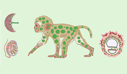
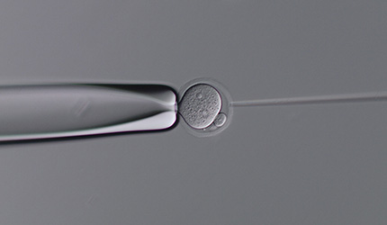
A recent study published in Cell has reported the generation of a live-birth chimeric monkey using a high contribution of embryonic stem cells (ESCs). This research was completed by Dr. LIU Zhen’s research team and Dr. SUN Qiang’s research team , and by Dr. Miguel A. Esteban’s research team.
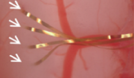
Dr. LI Xue’s lab, developed a hyperflexible electrode array for long-term recording and decoding of intraspinal neural activities. This work was recently published on Advanced Science.
A recent study published in Neuroscience Bulletin demonstrated that theta oscillations support prefrontal-hippocampal interactions in sequential working memory. This work was performed by researchers in Dr. YE Zheng’s Lab and Prof. ZHAN Shikun.

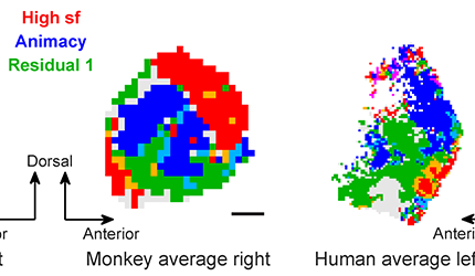
A recent study published in Nature Communications revealed the topographic organization of high-level visual areas in human and monkey brains. This work was performed by researchers in Dr. CHANG Le’s Lab. This work systematically investigated how visual features are represented and organized in the primate temporal lobe by combining functional MRI, electrophysiology, and deep learning, paving the way for the development of artificial visual systems with brain-like topographic organization.
A recent study published in Nature Metabolism demonstrated that astrocyte-specific overexpression of TMEM164 alleviating symptoms of neurodegenerative diseases in mouse models. This work was performed by the researchers in Dr. ZHOU Haibo’s Lab. This work has successfully identified transmembrane protein TMEM164 as a key regulator of neurotoxic astrocyte reactivity, and found that TMEM164 overexpression inhibited the induction of neurotoxic reactive astrocytes and neuronal death in both mouse and human astrocytes, as well as in Alzheimer’s disease (AD) and Parkinson’s disease (PD) mouse models. This discovery provides a potential therapeutic target for neurodegenerative diseases associated with neurotoxic reactive astrocytes.

The laboratory of Dr. XU Chun revealed neural mechanisms underlying associative learning of discontinuous events and demonstrated the critical role of the hippocampal CA1-Subiculum circuit in trace fear conditioning by combining behavioral analysis, optogenetics, and Calcium imaging.

A research team led by Dr.WANG Zuoren discovered that the activity of dopamine type 2 receptor-expressing neurons (Drd2+ neurons) in the dorsal bed nucleus of stria terminalis (dBNST) increased after long-term social isolation following weaning, leading to sex-specific abnormal behavior in male and female rats, by using techniques such as patch-clamp electrophysiology, chemogenetics manipulation, in vivo pharmacology experiments, and behavioral analysis. This study elucidated the involvement of dopamine type 1 and type 2 receptor-positive neurons in the dBNST region in regulating anxiety-like behavior induced by long-term social isolation, providing potential therapeutic targets for emotional disorders caused by social isolation.

A recent study published in Cell successfully mapped the cell-type taxonomy in the macaque cortex and revealed the relationship between cell-type composition and various primate brain regions, by using the self-developed spatial transcriptome sequencing technology Stereo-seq and snRNA-seq technology. These findings provide a molecular and cellular basis for further investigation into neural circuits.

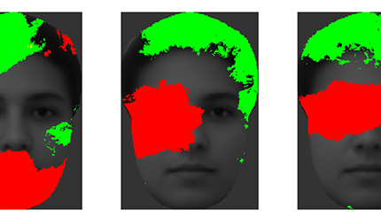
A recent study published in Cell Reports revealed the neural code for incomplete faces in the macaque face patches. This work was performed by researchers in Dr. CHANG Le’s Lab . This work systematically investigated how faces are represented in the primate brain when only a part of the face is shown, by conducting electrophysiological recordings in face-selective areas of the macaque inferotemporal cortex, paving the way for understanding the code of faces under natural conditions.
Chinese researchers led by Principal Investigator LIU Zhen, Principal Investigator SUN Qiang, and Dr. LU Zongyang, have develop an efficient gene integration method for rodent and primate embryos by MMEJ suppression. This gene knock-in method is referred to as CATI (CasRX-assisted Targeted Integration).
Recently, DU Jiulin’s research group, found the circadian regulation of synapse growth during neural development, taking the classic retinotectal synapse in zebrafish as an in vivo model implemented with the long-term time-lapse two-photon imaging. This study not only provides an important theoretical basis for the function of the circadian clock in regulating neural development, but also an important experimental basis for understanding the developmental rules of neuronal circuit formation.

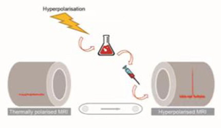
On June 1st, the journal Nature Chemistry published the Perspective article entitled: “Opportunities and challenges with hyperpolarized bioresponsive probes for functional imaging using magnetic resonance”. This is a collaborative work of Dr. Goran Angelovski from the International Center for Primate Brain Research, Dr. Ben J. Tickner and Dr. Gaoji Wang.
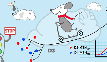
A recent study published in Neuroscience Bulletin reveals that the secondary motor cortex to dorsal striatum pathway is crucial for suppressing inappropriate responses in visual perceptual decision behavior. This work was performed by researchers in Dr. YAO Haishan’s Lab.
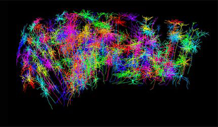
A new study published in Nature Neuroscience provides a comprehensive survey of the diversity and morphological types of dendrites of projection neurons in prefrontal cortex, by reconstructing and analyzing the complete morphology of dendrites and axons of nearly 2,000 single neurons in prefrontal cortex. This work reveals the relationship between dendritic morphology and axonal projections, and reconstructs the connection network between projection neuron types in prefrontal cortex. This work provides important clues for the in-depth study of the function of prefrontal cortex, basis for neuron simulation, and inspiration for artificial intelligence.
On April 25, 2023, the journal Physics of Life Reviews published a review paper online entitled “Structure and Function in Artificial, Zebrafish and Human Neural Networks.” This study was a collaborative effort between the DU Jiulin’s research group.
A new study published in Neuron revealed that cholecystokinin (CCK) neurons in the suprachiasmatic nucleus (SCN) play an important role in the regulation of the robustness of mammalian circadian clock. This study was performed by Dr. YAN Jun's lab.
A recent study published in Schizophrenia Bulletin demonstrated spatial and temporal abnormalities of spontaneous fixational saccades and their correlates with positive and cognitive symptoms in schizophrenia. This work was performed by researchers in Dr. WANG Wei’s lab and Dr. WANG Jijun’ team. This work suggests fixational saccades are a promising and easily obtainable biomarker for cognitive and positive symptoms and for complementary diagnosis in schizophrenia.

A recent study published in Genome Biology used SLIM-seq technology to map the dynamics of RNA m6A modification during maternal-to-zygotic transition (MZT) in mice, and revealed the functions of m6A reading and writing on mouse preimplantation embryonic development through multi-omics analysis and CRISPR/Cas13d-mediated gene knockdown.
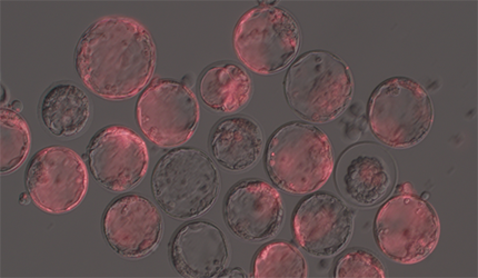
A recent study published in Cell Stem Cell reported the generation of monkey blastoids using na?ve ESCs and optimized protocol. In-vitro cultured monkey blastoids recapitulate gastrulation-like stage to germ layer formation. In-vivo transplantation of monkey blastoids triggers early pregnancy with gestation sacs. This study developed a potentially valuable system in investigating primate embryonic development.