Electrophysiology recording, as the ‘golden standard’ tools in the neuroscience, affords the advantages of high temporal resolution detection of the neural signal. However, the conventional neural interface has been limited by its instability caused by the mechanical mismatch between the rigid electrode and the soft brain tissue. The development of the ultraflexible neural interface has enabled a glial scare free chronic stable multichannel recording in the rodent model for over many months. Moreover, the combination of the ultraflexible neural interface with other advanced imaging methods has enabled new possibilities in the neuroscience study. And the application of the electrode in the spinal cord and peripheral nervous system potentially open new opportunities for clinical advances. Our group is focusing on the development of neural interfaces in both central and peripheral nervous systems.
1.Combining electrophysiological recording with neural imaging.
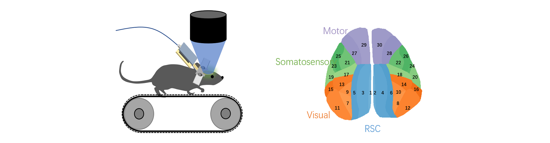
The brain is a massively interconnected network that spans across diverse spatial and temporal scales. In the past, neuroscientists are primarily working on either the microscale neural network in a small brain region or the macroscale functional connectivity across regions. Platforms that can link the functional connectivity across different spatial and temporal scales are currently missing, plus there is usually a lack of subcortical coverage. By combining large-scale ultraflexible electrode array with advanced cranial window technology, we enabled the possibility to study the functional connectivity between individual neurons to the whole cortex, with single-neuron resolution, at a time scale ranging from milliseconds to months. We are also working on combining electrical recording with more imaging technics and methods such as mini-scope and fMRI.
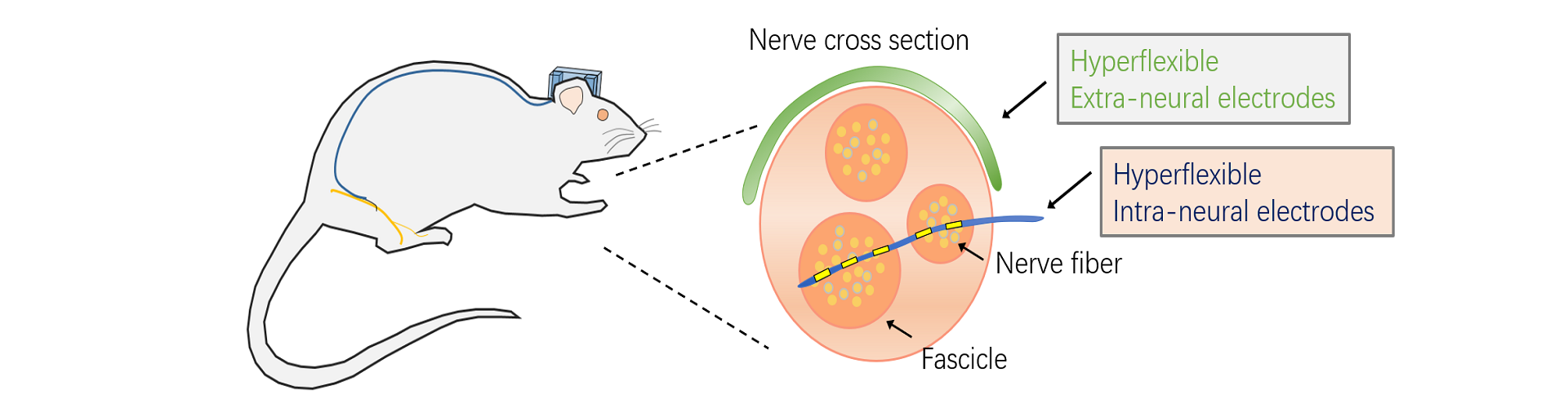
2.The development of ultraflexible peripheral neural electrodes.
The functional prosthesis can help patients with paralysis to regain parts of their motor and sensory functions. Currently, the most popular prosthesis is still based on EMG-controlled techniques, which only allows for movements with a single degree of freedom (DoF) at a time. However, the human hand has 21 DoFs. To create a real-time, robust, multiple DoFs prosthesis which can function naturally as human hands, a peripheral electrode which has multichannel, high selectivity, and chronic recording stability is crucial. We had been working on applying the concept of reducing probe size and flexibility to minimize the tissue response in the peripheral nervous systems. The preliminary results confirmed a minor tissue response near the implanted site in the rat sciatic nerve model. We are focusing on the development of a peripheral neural interface that can support chronic recording and stimulation. We are planning to apply the electrodes in clinically relevant models such as bladder dysfunction, epilepsy and depression.
3.The development of a close-loop and self-actuating brain machine interface (BMI).
After getting a chronically stable, high selectivity peripheral nerve electrode. I plan to implement the electrode into a close loop brain machine interface. There is some successful attempt trying to connect the Utah array based brain machine interface with muscle stimulator to help patients regain their upper limb motor function. However, the selectivity of the muscle stimulator is not high enough to let the patients perform natural behavior. And without sensory feedback, it is hard to control the force applied on the object. A flexible electrode based peripheral nerve interface that can achieve chronic recording and stimulation would be ideal for this application. This method not only minimizes the surgical invasiveness, increases the selectivity, but also includes natural sensory feedback.

Young Investigator