Understanding the functional architecture of the brain is one of the most interesting, but also challenging task of our times. This challenge is largely due to the extraordinary complicated structures and functional states of the brain, and the very limited access to them with our current research techniques. Our laboratory is interested in addressing such questions by developing new optical tools for brain research. Optical tools are the most promising way to observe and control neural activities in living organisms at micrometer or even nanometer scale noninvasively. By making use of this unique feature, we are going to develop the following new optical imaging techniques to obtain structural and functional information of the brain neural network and try to open new windows for brain research:
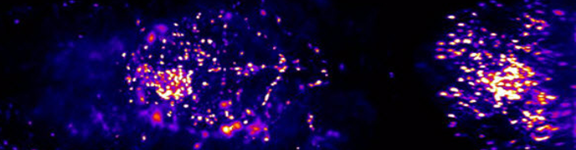
1. High resolution imaging and reconstruction of whole brain neural network
A complete map of the neural network is vital for fundamental understanding of the central nervous system. To achieve this goal, there have been numerous efforts using optical and electron microscopes to image brains of fruit flies and mice. Compared to electron microscopy, optical imaging has many advantages, such as high-speed, high penetration depth and compatibility with flexible fluorescence labeling. By making use of these advantages, we are going to develop new optical imaging methods, such as adaptive optics and super-resolution imaging techniques, and combine with advanced labeling techniques and tissue clearing techniques to imaging the entire brain of different animal models. Additionally, we will also develop new methods that arecompatible with the developed imaging methods to reconstruct the whole neural network reliably at a high speed. These techniques can be widely applied in brain science studies.
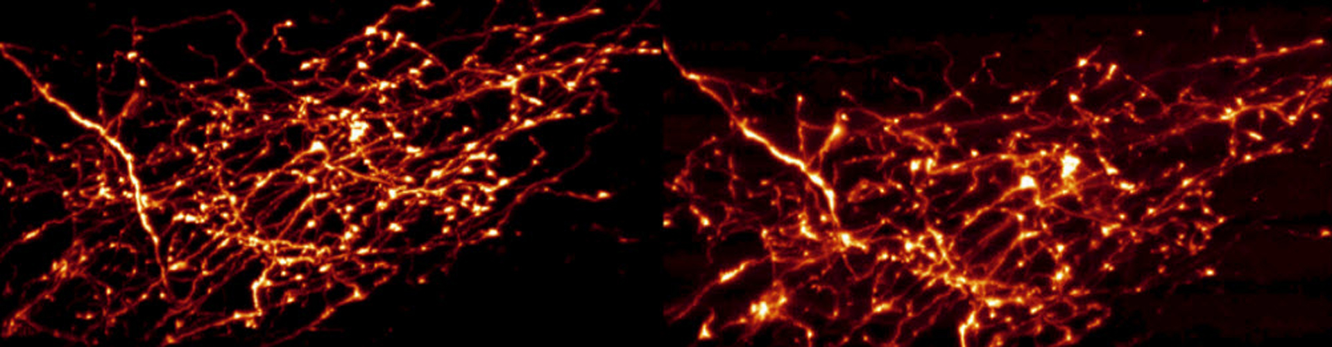
2. Deep brain optical imaging of neuronal activities with high resolution
Optical imaging can provide subcellular details of neurons at a high resolution, but has the drawback of limited penetration depth. This is due to the non-uniform optical properties of the biological tissue. The heterogeneity of the biological tissue will bend/scatter the light in unexpected ways and degrade imaging quality with increasing imaging depth. Currently, several techniques are developed to extend the penetration depth, such as two-photon imaging, three-photon imaging and adaptive optical microscope. We are interested in developing new ways to further extend the imaging depth in the brain of living or even awake animals. We hope to record functional activities on individual neurons or even dendrites, axons and synapses deep in the intact brain and open the new window for brain science studies.
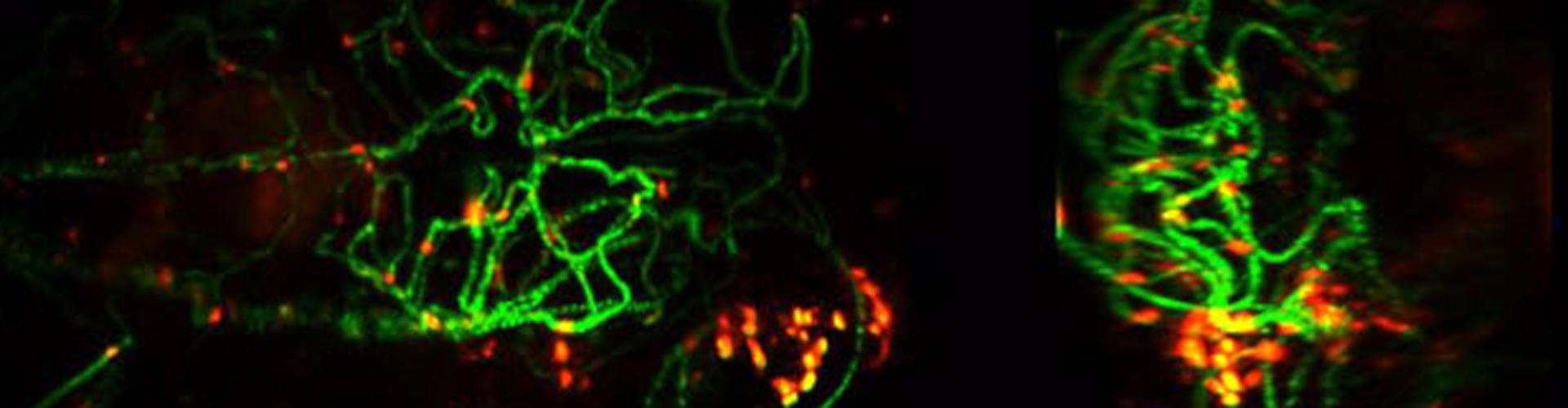
3. High-speed volume optical imaging of neuronal activities
Ideally, we want to get complete sets of information of the neuronal network activities in the brain, including all inputs and outputs of different neuronal circuits, so that we can analyze and hopefully understand how they work. However, current imaging techniques are either based on point-by-point or plane-by-plane scanning methods, and not fast enough to record activities of large volumes of neurons at the same time. To address this problem, we are interested in developing new 3D volume optical imaging techniques to record large groups of neuron’s activities at a high speed in awake animals. Such technique can pave the way to understandingneuronal activities of the whole brain.

Investigator